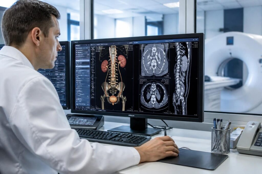
The uroscanner is an essential medical imaging examination for precisely visualizing the urinary system, including the kidneys, ureters, and bladder. Used in several clinical situations such as lower back pain, the presence of kidney stones, or recurrent urinary infections, it offers detailed images thanks to the use of X-rays combined with computer processing. Discover in which cases this abdominal scanner is truly indispensable and how it can guide an appropriate diagnosis and follow-up in urology.
The article in brief
The uroscanner facilitates the precise diagnosis of urinary disorders through advanced and rapid imaging.
- Comprehensive exploration of the urinary system: Precise visualization of the kidneys, ureters, and bladder
- Indicated for stones and lower back pain: Diagnosis of renal colic and hematuria
- Use in recurrent urinary infections: Investigation of underlying causes
- Reliable and safe examination: Preparation, procedure, and precautions to know
A key tool to better understand and treat common and complex urological pathologies.
Uroscanner: an essential tool in urological medical imaging
The uroscanner, or uro-computed tomography, is an imaging technique based on the controlled emission of X-rays to examine the urinary system. It allows capturing several hundred thin-slice images, then reconstructed in 2D or 3D by a computer. This method provides an overview that goes beyond a simple ultrasound, notably thanks to the possibility of injecting an iodinated contrast agent, which improves the visibility of structures such as the kidneys, ureters, and bladder. Its use has become common both in hospital centers and radiology offices in the city.
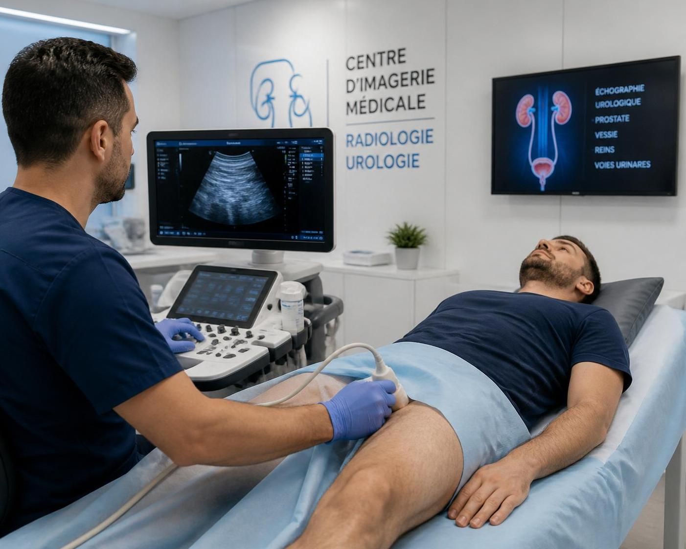
Various indications for a precise diagnosis
The uroscanner is essential when faced with symptoms suggestive of various urological disorders. Sudden lower back pain, often caused by the presence of kidney stones, requires this type of examination when ultrasound does not detect these stones sufficiently. The presence of blood in the urine (hematuria) or repeated urinary infections are also common reasons for prescription. Moreover, for patients with congenital malformations, kidney tumors, bladder, or urinary tract tumors, the uroscanner allows precise follow-up and a complete assessment to guide treatments. In the case of urological trauma, following a shock or accident, this fast and detailed examination is indispensable to assess lesions.
How does a uroscanner procedure take place?
The patient lies comfortably on a table that slowly moves through an open ring emitting X-rays. The examination generally lasts between 10 and 15 minutes. When a contrast product is required, it is injected intravenously into the elbow fold. This product enhances tissue distinction and notably allows a more precise observation of the excretory pathways. A diuretic injection may also be administered to better dilate these ducts. During image acquisition, the patient is asked to remain still and hold their breath for a few seconds at each scanner pass. Throughout the examination, communication with the medical team is maintained via an intercom.
List of the main indications for the uroscanner
- Renal colic: Search and localization of kidney or ureteral stones
- Unexplained hematuria: Identify the source of blood in the urine
- Recurrent urinary infections: Diagnose an anatomical or lithiasic underlying cause
- Tumor suspicions: Assessment and follow-up of renal, bladder, or excretory tract tumors
- Urological trauma: Lesion evaluation after accident or shock
- Congenital malformations: Detailed visualization of urinary morphology
Preparations and precautions to consider
Before a uroscanner with contrast product injection, a blood test is necessary to check kidney function, particularly creatinine levels. This step is crucial to reduce the risk of nephrotoxicity. Diabetic patients or those with renal insufficiency must inform their doctor. In the presence of allergic history to iodinated products, tests may be performed to prevent any risk of severe reaction. Contrary to some beliefs, fasting is not mandatory, but proper hydration is important. The bladder must be moderately filled to optimize images. However, the uroscanner should be avoided during pregnancy except in emergency situations, where the benefit-risk ratio is carefully assessed.
Possible risks and side effects
The examination is painless and the open ring of the scanner limits claustrophobia effects. Some sensations, such as transient warmth or a metallic taste, may occur at the time of injection. The diuretic usually causes an increased urge to urinate after the examination. The doses of radiation used are low, but repeated exposure should be avoided to limit potential long-term risks. The doctor remains vigilant regarding the use of the contrast agent, always evaluating the balance between diagnostic benefits and individual risks.
Summary table of advantages and disadvantages of the uroscanner
| Advantages | Disadvantages |
|---|---|
| Highly detailed 2D and 3D images of the urinary system | Use of X-rays involving exposure to radiation |
| Precise diagnosis for stones, tumors, and infections | Injection of contrast product can cause allergic reactions |
| Fast examination, feasible in emergencies | Not recommended during pregnancy without emergency |
| Well tolerated even by claustrophobic patients | Requires normal kidney function for iodinated product |
Professionals involved in image interpretation
The radiologist is the primary reference for interpreting the images obtained during the uroscanner. They can then direct to specialists such as the urologist for therapeutic management, the nephrologist in case of kidney disease, or the oncologist if cancer is suspected. Collaboration between these professionals ensures precise analysis and treatment adapted to the results. A second radiological opinion may be requested to strengthen diagnostic certainty.
What are the main indications of a uroscanner?
The uroscanner is prescribed in cases of lower back pain, suspected kidney stones, hematuria, recurrent urinary infections, malformations, or tumors of the urinary system.
Is fasting required before a uroscanner?
It is not systematically necessary to fast, but it is important to be well hydrated. The bladder should be moderately filled for better imaging quality.
What are the risks associated with injection of contrast product?
The contrast product may cause allergic reactions, mild or severe, and is contraindicated in case of significant renal insufficiency without particular monitoring.
Can a uroscanner be performed on pregnant women?
The uroscanner is generally avoided in pregnant women except in emergencies where the benefit for the mother outweighs the risks to the fetus.
How long does a uroscanner take?
The examination lasts between 10 and 15 minutes, depending on the need or not for injection of contrast product and the area to be examined.


